
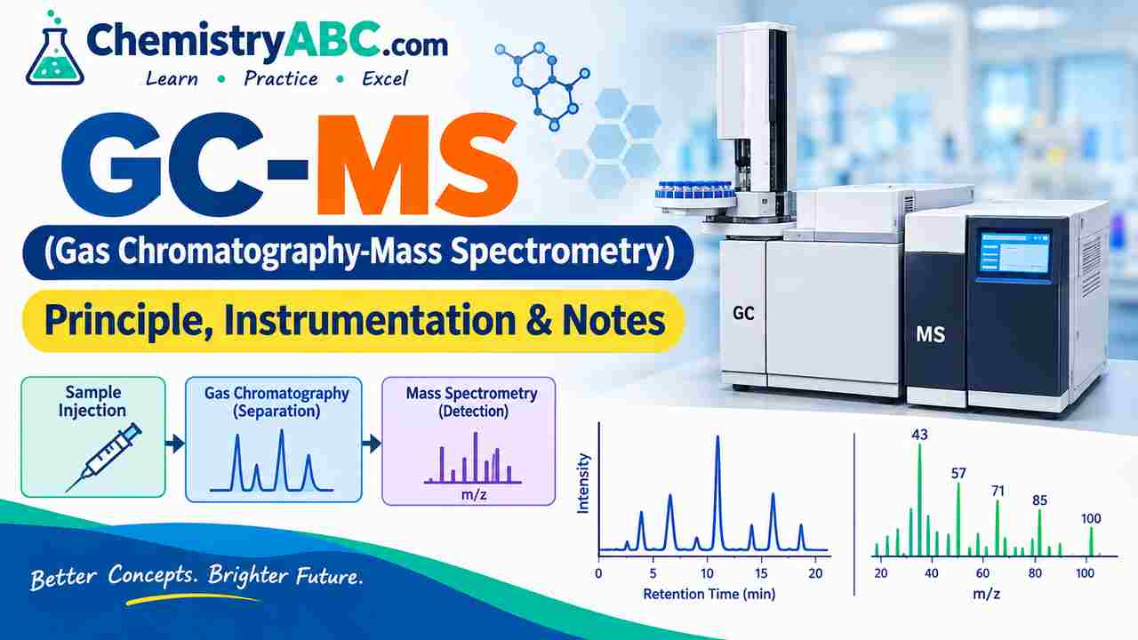
GC-MS (Gas Chromatography-Mass Spectrometry): Principle, Working & Notes
Explore the gold standard of hyphenated analytical techniques. Master GC-MS working principles, carrier gases, capillary columns, Electron Ionization (EI) vs. Chemical Ionization (CI), Quadrupole analyzers, and key mass fragmentation mechanisms.
- 1. What is GC-MS? (Fundamental Principle of Hyphenation)
- 2. Detailed Instrumentation: Architecture & Flowchart
- 3. Ionization Techniques: Electron Ionization (EI) vs. Chemical Ionization (CI)
- 4. Mass Analyzers in GC-MS: Quadrupole, TOF & Ion Trap
- 5. Key Mass Spectral Fragmentation Rules (McLafferty & Nitrogen Rule)
- 6. Solved Analytical Chemistry Practice Problems
- 7. Major Industrial, Environmental & Forensic Applications
- 8. Frequently Asked Questions (FAQs)
🔬 1. What is GC-MS? (Fundamental Principle of Hyphenation)
Gas Chromatography-Mass Spectrometry (GC-MS) is a powerful hyphenated analytical technique that combines the exceptional separation capability of Gas Chromatography (GC) with the precise structural elucidation and mass detection of Mass Spectrometry (MS).
1. GC Stage (Separation): Volatile, thermally stable analytes partition between a mobile carrier gas phase and a stationary liquid/polymer phase, separating based on boiling point and polarity (Retention Time, tR).
2. MS Stage (Identification & Quantification): Separated eluent molecules are ionized, fragmented into characteristic gas-phase ions, sorted by their mass-to-charge ratio (m/z), and detected to generate a unique molecular fingerprint.
⚙️ 2. GC-MS Instrumentation: Step-by-Step Architecture
The mobile phase carrier gas must be inert, ultra-high purity (>99.999%), and chemically non-reactive:
| Carrier Gas | Optimum Linear Velocity | Key Advantages | Primary Limitations |
|---|---|---|---|
| Helium (He) | 30 – 40 cm/s | Most common in GC-MS; non-flammable, excellent safety, high chromatographic efficiency | High cost, global supply constraints |
| Hydrogen (H2) | 40 – 50 cm/s | Fastest run times, highest van Deemter efficiency, economical, renewable generator source | Flammable; can hydrogenate unsaturated bonds in ion source |
| Nitrogen (N2) | 10 – 15 cm/s | Very cheap, readily available | Narrow optimum velocity; slow separation speed, lower MS pumping efficiency |
⚡ 3. Ionization Techniques: Electron Ionization (EI) vs. Chemical Ionization (CI)
Once analytes enter the high-vacuum ion source, they must be converted into gaseous ions to be manipulated by electric and magnetic fields:
| Feature | Electron Ionization (EI) | Chemical Ionization (CI) |
|---|---|---|
| Ionization Energy / Mechanism | Bombardment with high-energy electrons (standard 70 eV): M + e− → M+• + 2e− | Reagent gas (CH4, NH3, isobutane) is ionized first, reacting via proton transfer: M + [CH5]+ → [M + H]+ + CH4 |
| Type of Technique | Hard Ionization (Excess energy deposited into molecule) | Soft Ionization (Gentle proton transfer or hydride abstraction) |
| Molecular Ion Peak (M+•) | Often weak or completely absent due to extensive bond fragmentation | Prominent pseudo-molecular ion peak [M + H]+ or [M + NH4]+ |
| Spectral Library Matching | Universal reproducibility; directly searchable against standard NIST & Wiley Mass Spectral Databases | Reagent-gas dependent; requires customized reference standards |
| Primary Application | Structural elucidation, compound identification, fingerprint matching | Accurate determination of exact molecular weight (MW) |
🔍 4. Mass Analyzers in GC-MS Systems
The mass analyzer filters ions based on their mass-to-charge ratio (m/z). Common configurations include:
| Mass Analyzer | Operating Principle | Resolving Power | Major Practical Strengths |
|---|---|---|---|
| Quadrupole (Q) | Four parallel cylindrical rods with oscillating radiofrequency (RF) and direct current (DC) voltages; only ions of a specific m/z maintain stable trajectories to the detector. | Unit Mass Resolution (~1 amu) | Extremely robust, compact, linear quantitative dynamic range, highly cost-effective for routine testing. |
| Time-of-Flight (TOF) | Ions are accelerated with equal kinetic energy into a field-free drift flight tube; lighter ions travel faster and arrive at the detector earlier: t ∝ √(m/z). | High Resolution (HRMS: 20,000 – 60,000+) | Ultra-fast spectral acquisition speed (>500 spectra/sec); indispensable for comprehensive 2D Gas Chromatography (GC×GC). |
| Triple Quadrupole (QqQ) | Tandem MS/MS configuration where Q1 selects the precursor ion, Q2 serves as a collision cell (CID), and Q3 detects specific product fragments (MRM / SRM mode). | Unit Resolution with ultra-high selectivity | Eliminates matrix interferences; ultimate standard for trace level pesticide, drug, and environmental residue quantification. |
📐 5. Key Mass Spectral Fragmentation Rules
One of the most frequently tested fragmentation pathways in CSIR NET and GATE. Occurs in carbonyl compounds (aldehydes, ketones, esters, carboxylic acids) containing a γ-hydrogen atom:
• Example: Butyraldehyde (CH3CH2CH2CHO, MW = 72) undergoes McLafferty rearrangement to eliminate ethylene (C2H4, 28 amu), producing a diagnostic base peak at m/z = 44 [CH2=CH−OH]+•.
• A neutral organic molecule with zero or an even number of Nitrogen atoms has an EVEN molecular mass (M+•).
| Element | Stable Isotopes & Natural Abundance | Mass Spectral Diagnostic Signature |
|---|---|---|
| Chlorine (Cl) | 35Cl (75.8%) & 37Cl (24.2%) | Doublet peak separated by 2 amu: [M] and [M+2] in an intensity ratio of 3 : 1 |
| Bromine (Br) | 79Br (50.7%) & 81Br (49.3%) | Doublet peak separated by 2 amu: [M] and [M+2] in equal intensity (1 : 1) |
| Sulfur (S) | 32S (95.0%) & 34S (4.2%) | Notable [M+2] peak of approximately 4.4% height of [M] |
| Carbon (C) | 12C (98.9%) & 13C (1.1%) | [M+1] peak intensity ≈ 1.1% × Number of Carbon atoms |
💡 6. Solved Benchmark Analytical Problems
Problem 1: In the 70 eV EI mass spectrum of an organic compound, the molecular ion appears at m/z = 112 with an [M+2] peak at m/z = 114 having an intensity ratio of 3:1. The base peak appears at m/z = 77. Identify the compound and deduce the fragment ion at m/z = 77.
Step-by-Step Analytical Solution:
- Step 1: Isotope Peak Analysis: The 3:1 intensity ratio between [M] (112) and [M+2] (114) unequivocally confirms the presence of one Chlorine atom (35Cl / 37Cl).
- Step 2: Nitrogen Rule Check: Molecular weight m/z = 112 is an even integer, indicating either 0 or an even number of nitrogen atoms.
- Step 3: Radical Fragment Deduction: Subtracting the mass of 35Cl:
112 − 35 = 77. The remaining organic radical has a mass of 77. - Step 4: Base Peak Structure: m/z = 77 corresponds to the highly stable phenyl cation [C6H5]+ (6 × 12 + 5 = 77).
- Conclusion: The unknown analyte is Chlorobenzene (C6H5Cl). Loss of the chlorine radical (−•Cl) yields the phenyl base peak at m/z = 77.
Problem 2: Explain why pentan-2-one and pentan-3-one can be distinguished unambiguously by GC-MS under Electron Ionization.
Solution & Mechanistic Pathways:
- Both isomers share the same molecular formula (C5H10O, MW = 86), but exhibit distinctly different fragmentation pathways:
- Pentan-2-one (CH3COCH2CH2CH3): Contains γ-hydrogens on the propyl chain. It undergoes efficient McLafferty rearrangement, eliminating ethylene (28 amu) to yield a strong diagnostic peak at m/z = 58 [CH2=C(OH)CH3]+•. It also exhibits α-cleavage peaks at m/z = 43 [CH3CO]+ and m/z = 71.
- Pentan-3-one (CH3CH2COCH2CH3): Has only β-hydrogens, with no γ-hydrogens. Therefore, it cannot undergo McLafferty rearrangement. Its spectrum is dominated by simple α-cleavage, losing an ethyl radical (−C2H5) to give a base peak at m/z = 57 [CH3CH2CO]+.
- Conclusion: The presence of a prominent peak at m/z = 58 definitively confirms pentan-2-one.
🌐 7. Major Modern Applications of GC-MS
- Environmental Monitoring: Detection and quantification of volatile organic compounds (VOCs), polycyclic aromatic hydrocarbons (PAHs), dioxins, furans, and trace organochlorine pesticides in water and soil.
- Forensic Sciences: Identification of illicit drugs, narcotics, poisons, trace explosive residues, and arson accelerants at crime scenes.
- Food & Beverage Chemistry: Profiling essential oils, aromas, beverage contaminants, and verifying authenticity (e.g., wine and olive oil adulteration).
- Pharmaceuticals & Metabolomics: Tracking residual solvents, impurity profiling, and clinical screening of inborn metabolic disorders via organic acid profiling in urine.
❓ 8. Frequently Asked Questions (FAQs)
No. GC-MS requires samples to be volatile and thermally stable at temperatures up to 300 °C. Non-volatile compounds with polar functional groups (such as amino acids, fatty acids, or carbohydrates) must first undergo chemical derivatization (e.g., silylation using BSTFA/TMCS, methylation, or trifluoroacetylation) or be analyzed using LC-MS (Liquid Chromatography-Mass Spectrometry).
A Total Ion Chromatogram (TIC) represents the sum of all ion intensities across the scanned mass range plotted against retention time, showing all components in the sample. An Extracted Ion Chromatogram (EIC) isolates and plots only a single specific m/z value, drastically improving signal-to-noise ratio and selectivity for targeted peak quantification.
At 70 eV, the de Broglie wavelength of ionizing electrons (~0.14 nm) closely matches typical molecular bond lengths, maximizing ionization efficiency and cross-section. Crucially, 70 eV spectra are highly reproducible across different instrument manufacturers, enabling universal search against the NIST and Wiley reference libraries.
Educational Disclaimer: ChemistryABC.com provides educational resources and analytical chemistry guides strictly for self-study and university examination preparation. All product names, trademarks, and registered trademarks belong to their respective owners.
For academic queries, feedback, or suggestions, please write to us at: [email protected]